
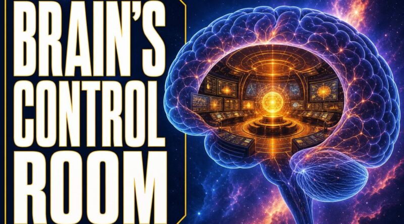
Brain’s Hidden Control Room Finally Mapped
Brain’s Hidden Control Room Finally Mapped
There’s a structure inside your skull the size of your thumb. It controls every breath you take, every heartbeat, every moment you fall asleep and wake up again. Without it, you’d be gone in minutes. And until July 2026, science had never truly seen inside it — not at the level of individual cells.
That changed when researchers at IIT Madras built something called Anchor. And what they found rewired what we thought we knew about the human brain.
The Part of Your Brain Nobody Talks About
Most people, when they think about the brain, picture the wrinkled outer layer — the cortex. That’s the part linked to memory, language, personality. It gets all the attention.
But deep inside, sitting at the base of the brain where it connects to the spinal cord, is the brainstem. No dramatic folds. No glamour. Just a dense, compact structure that quietly runs the machinery keeping you alive.
the brainstem controls breathing. It regulates your heartbeat. It decides when you sleep and when you wake. It acts as the main cable between your brain and the rest of your body — every signal travelling up or down passes through it. Damage it severely, and no amount of advanced medicine brings you back.
Here’s the strange part: for all its importance, scientists had never produced a complete, cell-level map of the human brainstem. We had rough outlines. We had partial pictures. But the full, three-dimensional picture — showing exactly which cells live where, which chemical systems they belong to, how the nerve pathways are arranged — that didn’t exist.
Until now.
What Anchor Actually Is
On 13 July 2026, scientists at the Sudha Gopalakrishnan Brain Centre at the Indian Institute of Technology Madras announced something they’d spent years building: the world’s most detailed three-dimensional atlas of the human brainstem at cellular resolution.
They named it Anchor — short for Atlas of Neurochemical Characterisation of the Human Brainstem with 3D Reconstruction.
The scale of the project is worth sitting with. The team assembled more than 500 tissue sections, taken from foetal brains, childhood brains, and adult brains. Each section was examined, chemically tagged, and then reconstructed digitally into a single, unified three-dimensional map.
To tell different cell types apart, they used eight distinct chemical markers — each one acting like a different coloured highlighter on a different population of cells. The result: Anchor identifies more than 200 separate clusters of brain cells and nerve pathways inside the brainstem alone.
Two hundred clusters. In a structure the size of your thumb.
That number alone tells you something important: the brainstem is not a simple relay switch. It is an extraordinarily complex neighbourhood, packed with distinct communities of cells doing very different jobs — and we are only now beginning to see who lives where.
Why 86 Billion Neurons Weren’t Enough to Understand This
The human brain contains roughly 86 billion neurons. That number gets quoted often, usually to make the brain sound impressively large. But raw neuron count tells you almost nothing about how the brain actually works.
Understanding the brain requires knowing which neurons are where, what chemical systems they use to communicate, and how they’re wired to each other. That’s the difference between knowing a city has 10 million people and actually having a street map.
For the cortex — the outer brain — researchers have been building those street maps for decades. Imaging technology, surgical studies, and decades of neuroscience have given us reasonable maps of the cortex’s major regions.
The brainstem was different. Its density, its complexity, and the technical difficulty of studying it at cellular resolution meant it remained, in a real sense, unmapped. Scientists knew what the brainstem did — they’d known that for generations. What they didn’t have was a precise picture of the machinery doing it.
That gap mattered enormously. Conditions including Parkinson’s disease, sleep disorders, and certain forms of paralysis all involve brainstem circuits. Treating them more precisely requires knowing exactly which cell populations are affected. Without a detailed map, researchers were working partly in the dark.
Anchor is the map they didn’t have.
How You Build a 3D Atlas of the Human Brain’s Most Complex Structure
The process behind Anchor is worth understanding, because it reveals just how hard this kind of science actually is.
The team at IIT Madras’s Sudha Gopalakrishnan Brain Centre started with physical tissue — actual sections of human brainstem tissue collected across different life stages: foetal, childhood, and adult. Using tissue from multiple stages was deliberate. The brainstem changes as we develop, and a complete atlas needed to capture that variation, not flatten it.
Each of the 500-plus tissue sections was chemically stained using eight different markers. Think of each marker as a key that fits a specific lock — only certain cell types carry that lock, so only those cells light up under that marker. By stacking multiple markers, researchers could distinguish cell populations that would otherwise look identical under a microscope.
Then came the reconstruction: taking hundreds of flat tissue slices and computationally assembling them into a single three-dimensional structure. That’s not just a technical challenge — it requires the kind of precision where a single misaligned section can distort the entire map.
The output was Anchor: 200-plus identified clusters, mapped in three dimensions, covering the full developmental range of the human brainstem. Nothing at this resolution had existed before.
What This Changes — And Why It Matters Right Now
A map is only useful if you use it. So what does Anchor actually unlock?
The most immediate application is in understanding neurological conditions that originate in the brainstem. Parkinson’s disease, for instance, involves the progressive loss of specific cell populations — many of them located in brainstem regions. Knowing precisely where those populations sit, what chemical markers they carry, and how they connect to other circuits gives researchers a far more precise target for both diagnosis and treatment.
Sleep disorders are another area. The brainstem is central to regulating sleep and wakefulness. With 200-plus identified clusters now mapped, scientists can begin asking which specific populations are disrupted in conditions like sleep apnea or narcolepsy — and design interventions accordingly.
Beyond disease, Anchor matters for basic neuroscience. Every time researchers study a brainstem circuit in an animal model and want to know if the findings apply to humans, they need a human reference point. Anchor becomes that reference — a shared, detailed baseline that the global research community can use, compare against, and build on.
The fact that this came out of IIT Madras’s Sudha Gopalakrishnan Brain Centre is significant in its own right. It places India at the frontier of a field — human brain mapping — that has historically been dominated by institutions in the United States and Europe. Anchor isn’t a contribution to that conversation. It leads it.
Final Thought
The brainstem has been keeping humans alive for as long as humans have existed. Every breath taken by every person who ever lived ran through this structure. And yet, until 13 July 2026, we had never seen it clearly — not at the level of individual cells, not in three dimensions, not across the full arc of human development.
Anchor changes that. More than 500 tissue sections, eight chemical markers, and 200-plus identified clusters later, the Sudha Gopalakrishnan Brain Centre at IIT Madras has handed neuroscience something it has needed for decades: a real map of the human brain’s most essential structure.
The human brain contains 86 billion neurons. Anchor is the beginning of finally understanding what they’re all actually doing.
Frequently Asked Questions
What is the Anchor brain atlas and who created it?
Anchor is the world’s most detailed three-dimensional atlas of the human brainstem at cellular resolution, created by scientists at the Sudha Gopalakrishnan Brain Centre at IIT Madras and announced on 13 July 2026.
What does the brainstem do and why is it important?
The brainstem controls breathing, regulates heartbeat, and manages sleep-wake cycles. It also acts as the main communication cable between the brain and the rest of the body, making it essential for survival.
Has the human brainstem ever been fully mapped before?
No, scientists had only rough outlines and partial pictures before 2026. A complete, cell-level three-dimensional map showing which cells live where and how nerve pathways are arranged had never existed until the Anchor atlas was created.
Recommended Reading
Explore these hand-picked resources to dive deeper into this topic:
- Thinking, Fast and Slow by Daniel Kahneman
- The Brain That Changes Itself by Norman Doidge
- Brain Anatomy Model Kit (3D educational replica)
As an Amazon Associate, we earn from qualifying purchases. This helps support Fact Storm Hub at no extra cost to you.
Sources
- https://www.ovid.com/jnls/neur/fulltext/10.4103/0028-3886.253639~construction-of-indian-human-brain-atlas
- https://www.bbc.com/news/articles/cg53l737v1qo
- https://www.youtube.com/watch?v=pCikHutVj_Y
- https://pubmed.ncbi.nlm.nih.gov/30860125/
- https://iitm.humanbrain.in/
🤖 AI Content Disclosure
This article was created using AI-assisted research and writing tools, then reviewed for quality and accuracy. Facts are sourced from publicly available web research, but readers should verify critical information from primary sources.
Published for educational and entertainment purposes. Last reviewed: August 2026